
FRCA anatomy, drawn as clear schematics
97 anatomical and sonoanatomy schematics for anaesthesia: the relations, landmarks and pathways you need to understand, label and explain.
Subscriber? Sign in and open GAG from your course.
Anaesthetic anatomy diagrams you can redraw
Anatomy for anaesthesia is mostly about relations: which nerve runs where, what lies next to the needle, which level is which. GAG’s anatomy items are simplified, labelled schematics that make those relations clear. They are designed for understanding and recall, not as realistic illustrations.
Each item gives you:
- a labelled schematic, with a written Description of the visual;
- Reveal labels, one at a time, to practise identification and labelling;
- Explain in 60 seconds, and the clinical application where the item has it;
- links to related GAG items, such as the matching block or the guidance on safe vascular access.
Sonoanatomy, schematically
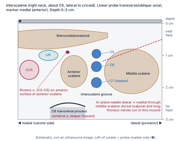
23 items are sonoanatomy schematics or combine anatomy with sonoanatomy: drawn ultrasound views with depth, orientation and the structures to find, for regional anaesthesia, vascular access, focused echo and lung ultrasound in pneumothorax. They are not ultrasound images and do not replace supervised scanning.
Anatomy matters in both parts of the exam: for the Primary practical exams in Primary Practical, and for regional anaesthesia and procedures in Final Practical.
Topics you will find in Anatomy
A selection of 33 published titles. The topic directory lists all 97.
Airway, larynx and neck
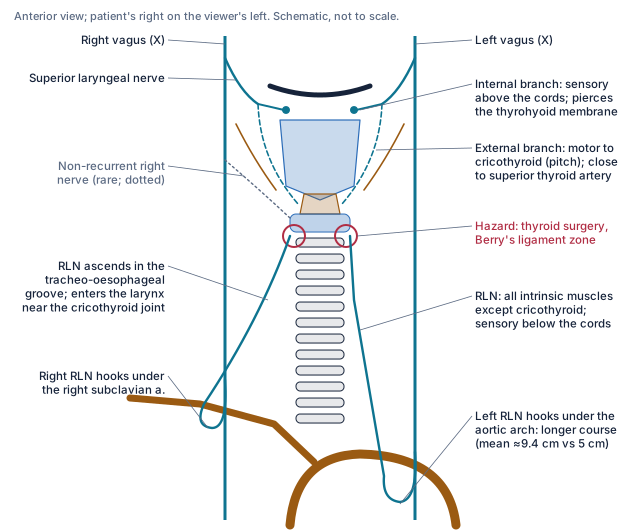
- Laryngeal innervation: superior and recurrent laryngeal nerves
- Larynx: cartilages, joints, muscles and glottis
- Front of neck: cricothyroid membrane, relations and ultrasound
- Head and neck surgical relations: thyroid, carotid and facial nerve
- Neck cross-section at C6: relations, fascia and hazards
Head, brain and spinal cord
- Skull base foramina and their contents
- Brain herniation, brainstem reflexes and focal lesion localisation
- Circle of Willis: arteries, territories and variants
- Spinal cord cross-section: tracts, crossings and lesion patterns
Plexuses and regional anaesthesia sonoanatomy
- Brachial plexus: roots to terminal branches, variants and injury patterns
- Cervical plexus blocks (superficial, intermediate, deep): sonoanatomy at C4
- Interscalene block window: sonoanatomy and the phrenic nerve
- Supraclavicular block window: subclavian artery, first rib and pleura
- Lumbar plexus: roots, psoas relations, branches and territories
- Ankle block: five nerves and their windows
Spinal nerves and the neuraxis
- Spinal nerve: roots, ganglion, rami and sympathetic connections
- Conus, dural sac and surface landmarks: lumbar neuraxis in sagittal view
- Neuraxial functional levels: sympathetic, cardioaccelerator and visceral pathways
- Epidural space: boundaries, contents and lumbar versus thoracic
- Neuraxial ultrasound: views and counting levels
Thorax and vascular access
- Thoracic inlet and first rib: relations, pleural dome and thoracic outlet
- Typical rib: features, joints, costal groove and pleural relations
- Pleura, recesses and the safe triangle for chest drains
- Internal jugular vein: course, relations and ultrasound-guided cannulation
- Central venous catheter tip position, malposition pathways and confirmation
Heart, focused echo and lung ultrasound
- Focused echo: parasternal long- and short-axis views
- Focused echo: apical four-chamber, subcostal and IVC views
- Lung ultrasound in pneumothorax: sliding, A-lines, barcode and lung point
- Congenital shunts, cyanotic physiology and Eisenmenger syndrome
- Left-sided valve lesions: anatomy and pressure–flow consequences
- Coronary arteries: origins, course, dominance and territories
Obstetric anatomy
- Aortocaval compression and uterine displacement
- Pregnancy: neuraxial and airway anatomical changes
What a GAG visual looks like
Frequently asked questions
Are these anatomical drawings or photographs?
Neither: they are simplified schematics. Each shows the structures and relations that matter for understanding and explaining a topic, and leaves out detail that does not. They are not to scale and are labelled as schematics.
Can GAG replace an anatomy atlas or ultrasound practice?
No. GAG helps you understand and recall key relations and explain them. Keep using atlases, prosection, hands-on ultrasound scanning and supervised clinical teaching for anatomy and regional anaesthesia.
Do the sonoanatomy schematics show real ultrasound images?
No. The sonoanatomy items are drawn schematics of the ultrasound view, showing what to identify and where, with depth and orientation marked. They are not ultrasound images.
How do I practise labelling?
Use Reveal labels on a labelled diagram: try naming each structure first, then reveal its label to check. Then sketch the diagram from memory and compare.
Continue: Graphs & Science · Guidelines & Tables · GAG overview
GAG comes with full access to every course
GAG is included in Primary Written, Primary Practical, Final Written and Final Practical, with exam-focus prompts for each.
The free preview does not include GAG.
Already registered? Sign in